· 高通量单细胞分析:同时生成上千单细胞测序数据 · 样品灵活性高:一次实验可同时对8个不同样品进行单细胞分析 · mRNA 3’端捕获测序:mRNA 3’端捕获测序差异表达分析所需reads数更少 · 减少样品处理操作:自动完成文库构建前的细胞分离、挑选及混样,偏差更小 · 兼容illumina平台:构建的文库支持illumina平台测序 |
|
■ 产品说明 |
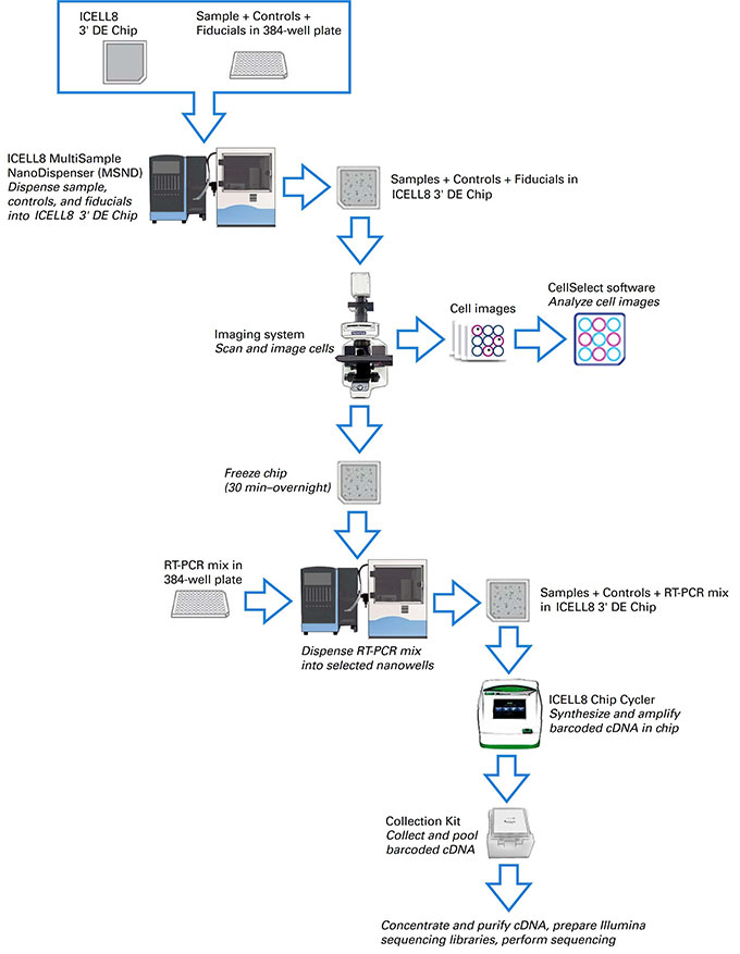
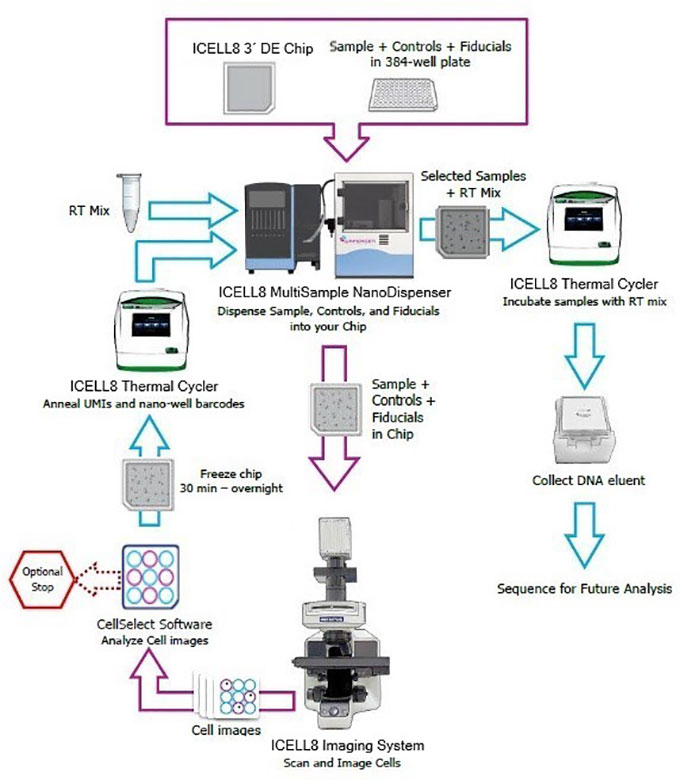
通过结合Takara Bio单细胞RNA-Seq文库构建的技术优势及ICELL8 Single-Cell System单细胞分选的优良性能,ICELL8 Chip and Reagent 3’DE Kit和ICELL8 3’DE for UMI Reagent Kit实验流程均可以一次制备最多八个不同样品的Illumina测序文库,进行不含UMI或含UMI的高通量单细胞基因表达差异。 样品细胞经过染色、稀释,使用ICELL8单细胞分选系统将其分注到微孔芯片ICELL8 3’DE Chip(Code No. 640143)中。微孔芯片表面排布5,184个纳升微孔,并预先固相化了Barcodes。通过单细胞分选系统的成像功能和配套的CellSelect软件,含有活单细胞的微孔将被挑选出来,并添加RT-PCR试剂,在芯片中完成cDNA合成与扩增。从芯片中将cDNA产物混合收集,使用基于转座酶的方法富集mRNA 3’ 端来源的cDNA,随后通过PCR扩增整合入Illumina index序列。 通过每个微孔特异的Barcode序列,能从最终的文库测序数据中回溯每个活的单细胞来源的数据,并可基于mRNA 3’ 端计数,进行差异基因表达分析。配合ICELL8单细胞分选系统,ICELL8 Chip and Reagent 3’DE Kit和ICELL8 3’DE for UMI Reagent Kit为高通量单细胞差异表达分析提供了高效、经济的解决方案,一次实验即可从上千个活的单细胞获得高质量3’ 端测序数据。 |
|
■ 产品组分 |
完整的工作流程需要下列Takara Bio产品。同时,请参考对应的User Manual得到一份完整的试剂耗材清单: |
用于ICELL8 Single-Cell System(不含UMI分子标签工作流程) |
· ICELL8 Chip and Reagent 3’DE Kit (Code No. 640164、640165、640166)* · ICELL8 384-Well Source Plate and Seal (5/pack) (Code No. 640192) |
User Manual:ICELL8® 3’DE Reagent Kit User Manual |
* NOTE:ICELL8 Chip and Reagent 3’DE Kit包含以下四种制品,640143、640167、640048、640109。640164包装量对应1张芯片用量,640165包装量对应3张芯片,640166包装量对应5张芯片。 |
· ICELL8 3’DE Chip (Code No. 640143) · ICELL8 3’DE Reagent Kit(Code No. 640167) · ICELL8 Collection Kit (Code No. 640048) · ICELL8 Loading Kit (Code No. 640109) |
|
用于ICELL8 Single-Cell System(含UMI分子标签工作流程) |
· ICELL8 3’DE Chip (Code No. 640143) · ICELL8 3’DE for UMI Reagent Kit(Code No. 640005) · ICELL8 Collection Kit (Code No. 640048) · ICELL8 Loading Kit (Code No. 640109) · ICELL8 384-Well Source Plate and Seal (5/pack) (Code No. 640192) |
User Manual:ICELL8® 3’DE for UMI Reagent Kit User Manual |
|
|
图1 ICELL8 Chip and Reagent 3’DE Kit工作流程 |
|
|
图2 ICELL8 3’DE for UMI Reagent Kit工作流程 |
|
参考文献 |
1. Ren, Z. et al. Single-Cell reconstruction of progression trajectory reveals intervention principles in pathological cardiac hypertrophy. Circulation, 141, 1704–1719 (2020) 2. Wang, L. et al. Single-cell reconstruction of the adult human heart during heart failure and recovery reveals the cellular landscape underlying cardiac function. Nat. Cell Biol. 22, 108–119 (2020) 3. Krieger, T. G., et al. Modeling glioblastoma invasion using human brain organoids and Single-Cell transcriptomics. Neuro-Oncology, 22, 1138–1149 (2020) 4. Yekelchyk, M., Guenther, S., Preussner, J. & Braun, T. Mono- and multi-nucleated ventricular cardiomyocytes constitute a transcriptionally homogenous cell population. Basic Res. Cardiol. 114, 36 (2019) |
|
产品详情请点击: |
|
|
|