上海金畔生物科技有限公司提供Annexin V-Alexa Fluor 647细胞凋亡检测试剂盒 ,欢迎访问官网了解更多产品信息。
| 产品编号 | BA00103 |
| 英文名称 | Annexin V-AF647 Apoptosis Detection Kit |
| 中文名称 | Annexin V-Alexa Fluor 647细胞凋亡检测试剂盒 |
| 别 名 | Annexin V-AF647/PI Apoptosis Detection Kit; Annexin V-AF647 Apoptosis Detection Kit; Annexin V-Alexa Fluor 647/PI Kit; Annexin V-Alexa Fluor 647/PI Kit; Annexin V-AF647/PI Kit; Annexin V-AF647/PI双染细胞凋亡检测试剂盒; Annexin V-AF647细胞凋亡检测试剂盒; AF647 Annexin V/PI细胞凋亡检测试剂盒; Annexin V-AF647/PI双染细胞凋亡检测试剂盒; ANNEXIN V- AF647凋亡检测试剂盒; Annexin V-AF647/PI细胞凋亡检测试剂盒; 细胞凋亡检测试剂盒; 细胞凋亡试剂盒; |
 |
Specific References (2) | BA00103 has been referenced in 2 publications.
[IF=10.164] Wang D et al. Loss of 4.1N in epithelial ovarian cancer results in EMT and matrix-detached cell death resistance.. Protein Cell. 2020 May 25. FCM ; Human.
PubMed:32448967
[IF=5.714] Xiangming Liu. et al. Taxifolin ameliorates cigarette smoke-induced chronic obstructive pulmonary disease via inhibiting inflammation and apoptosis. INT IMMUNOPHARMACOL. 2023 Feb;115:109577
PubMed:36584569
|
| 保存条件 | Store at 4℃. |
| 注意事项 | This product as supplied is intended for research use only, not for use in human, therapeutic or diagnostic applications. |
| 产品介绍 | 磷脂酰丝氨酸(PS)是一种带负电荷的磷脂,正常细胞中,PS只分布在细胞膜脂质双层的内侧,而在细胞凋亡早期,细胞膜 PS由脂膜内侧翻向细胞膜外侧,使PS暴露在细胞膜外表面。Annexin V是一种分子量为35~36kD 的Ca2+依赖性磷脂结合蛋白,与磷脂酰丝氨酸有高度亲和力,故可通过细胞外侧暴露的磷脂酰丝氨酸与凋亡早期细胞的胞膜结合。因此Annexin V被公认为检测细胞早期凋亡的灵敏指标之一。 将Annexin V进行Alexa Fluor 647标记,以标记了的Annexin V作为探针,利用荧光显微镜或流式细胞仪可检测细胞凋亡的发生。碘化丙啶(Propidium Iodide, PI)是一种核酸染料,它不能透过正常细胞或早期凋亡细胞的完整的细胞膜,但对凋亡中晚期的细胞和坏死细胞,PI能够透过细胞膜而使细胞核染红。因此采用Annexin V与PI双染的方法,就可以将处于不同凋亡时期的细胞区分开来。 |
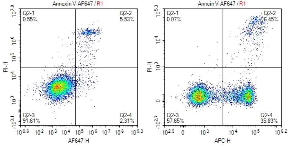
| 产品图片 |
 Jurkat cells (T-cell leukemia,human) treated with 10 μM of camptothecin for 4 hours(panel right) or untreated control(panel left).
1. Wash cells twice with cold PBS and then resuspend cells in 1×Binding Buffer at a concentration of 1×10⁶ cells/ml. 2. Transfer 100 μl of the solution (1×10⁵cells) to a 5 ml culture tube. 3. Add 5 μl of Annexin V-AF647. 4. Add 5 μl PI. 5. Gently vortex the cells and incubate for 15 min at RT (25°C) in the dark. 6. Add 400 μl of 1×Binding Buffer to each tube.Analyze by flow cytometry within 1 hr. |
